Rewriting Life
Intelligence Explained
Tracking and understanding the complex connections within the brain may finally reveal the neural secret of cognitive ability.


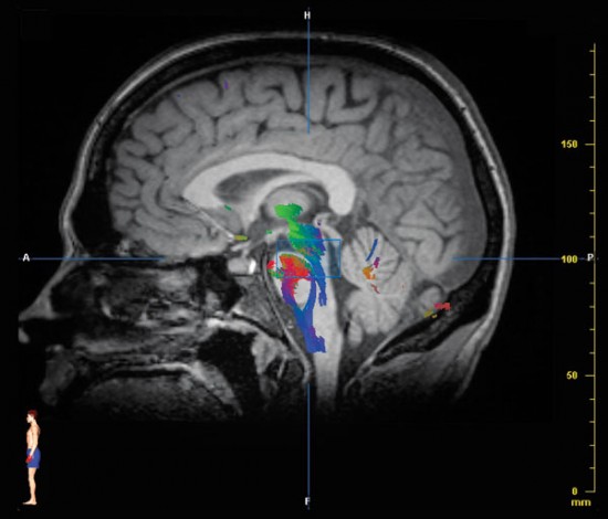
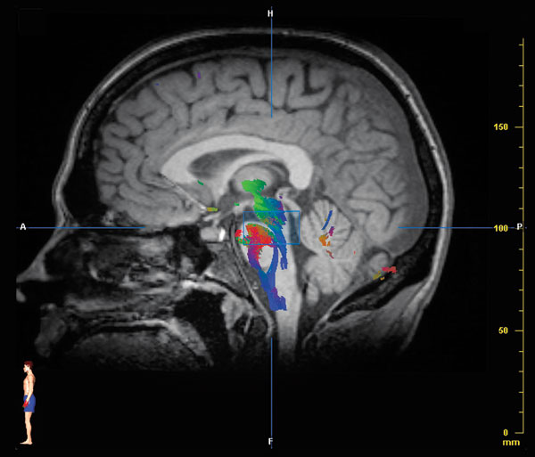
A series of black-and-white snapshots is splayed across the screen, each capturing a thin slice of my brain. The gray-scale pictures would look familiar to anyone who has seen a brain scan, but these images are different. Andrew Frew, a neuroscientist at the University of California, Los Angeles, uses a cursor to select a small square. Thin strands like spaghetti appear, representing the thousands of neural fibers passing through it. A few clicks of the cursor and Frew refines the tract of fibers pictured on the screen, highlighting first my optic nerve, then the fibers passing through a part of the brain that’s crucial for language, then the bundles of motor and sensory nerves that head down to the brain stem.
Frew is giving me a tour of my white matter–the tissue connecting the neurons, or nerve cells, that make up gray matter. Something about the twisting, turning neural wires that ferry information between the neurons–their individual thickness, perhaps, or their abundance, or the specific paths they take from one part of the brain to another–may explain, at least in part, the variations in human intelligence.
Scientists have been searching more than two centuries for the source of intelligence–the general cognitive ability often quantified in the form of IQ. With the advent of technologies such as magnetic resonance imaging (MRI), researchers concentrating mainly on gray matter have been able to map the parts of the brain that appear to play a role. But this has taken them only so far, and the focus on gray matter has not told the whole story. Not until the last few years, as new variations of MRI home in on the brain’s white matter, has a deeper understanding begun to emerge. “Scientists are now able to switch the focus from particular regions of the brain to the connections between those regions,” says Sherif Karama, a psychiatrist and a neuroscientist at McGill University’s Montreal Neurological Institute. Their initial findings have led Karama and others to believe that neural wiring and the way it carries information around the brain may be crucially important to IQ.
Until fairly recently, only a few scientists were studying how brain structure might be related to IQ, in part because the idea of a biological and genetic basis for intelligence has long been controversial. Since people from different ethnic groups often score differently on intelligence tests, such studies may raise contentions of racism, and critics fear potential abuses such as discrimination in education or employment. Nonetheless, new imaging techniques have allowed types of studies never before possible, and the number of research groups focusing on this question is growing quickly. Many of these groups are setting their sights on white matter.
The hope is that finding the brain areas and circuits involved in intelligence will provide new insight into neurological and psychiatric diseases that impair cognition, such as Alzheimer’s and schizophrenia. “If you want to understand cognitive decline, you need to understand how cognition is manifested and put together in the brain,” says Rex Jung, a neuroscientist at the Mind Research Network in Albuquerque, NM. The research may also improve understanding of learning disabilities such as dyslexia and ADHD, perhaps leading to better treatments. But other potential applications could be more controversial. Some scientists envision a day when brain scans are used to estimate IQ. Sandra F. Witelson, a neuroscientist at the Michael G. DeGroote School of Medicine at McMaster University in Ontario, says, “It’s not a wild guess to say that sometime in the future, brain scans will be part of a group of tools that try to indicate what level someone’s ability is going to be.”
Big Brains
Neuroscientist Paul Thompson is one of those researchers studying brain structure and IQ, but that wasn’t what he planned on when he started his lab at UCLA: he focused on the wave of changes in the brain that characterize Alzheimer’s and schizophrenia. Because serious cognitive deficits accompany both of those diseases, however, Thompson and his collaborators tested cognitive function in their subjects. When they began to look more closely for variables that correlated with brain structure, they found that intelligence seemed to be among the most significant. “IQ came in as a key factor that determines how the brain looks,” Thompson says.
Scientists who study intelligence typically define it in comparative terms, as a general cognitive ability measured against a mean. A quantifiable “general intelligence factor,” known as g, can be statistically extracted from scores on a battery of intelligence tests. While some people clearly have particular areas of talent, those who score well on one test are likely to score well on others as well, reflecting a higher g.
Researchers have yet to find a simple neural explanation for g. In 2001, Thompson showed that it is correlated with volume in the frontal cortex, a result consistent with a number of studies that have linked intelligence to overall brain size. But size is a crude measure: while larger brains may be smarter on average, it’s not clear if that’s because they have more nerve cells, more connections between cells, or more of the fibers that carry neural signals. Any of these factors can result in a larger brain or thicker cortex, but neither of these things is necessary for great intelligence. Studies of Albert Einstein’s brain, for example, have found that it was typical in size, or even a bit on the small side. (It was missing a wrinkle in the inferior parietal lobe, which is behind the frontal cortex; some have speculated that this quirk allowed the neurons in that region to communicate more effectively.)
As structural brain imaging has become more sophisticated, scientists have focused on sections of the brain involved in specific tasks, including sensory processing, memory, attention, and decision making. Different studies have connected different areas with intelligence, however, making it difficult to come to an overarching conclusion about its anatomical basis.
But what if the key to intelligence is neither an individual area of the brain nor its total volume but the network over which information is transmitted and integrated? In 2007, Jung and Richard Haier, now professor emeritus of psychology at the University of California, Irvine, developed the first comprehensive theory drawn from neuroimaging of how the brain gives rise to intelligence. Gathering information from 37 published papers that had used imaging to study intelligence, they mapped out the brain areas that had been pinpointed in at least a third of the studies to sketch a network of regions spanning the frontal and parietal lobes.
The network consists of about 10 nodes, or clusters of cells, that had been linked to attention, working memory, and facial recognition, among other cognitive functions. Applying existing theories of how information flows in the brain, Jung and Haier hypothesized that neural signals travel from nodes near the back of the brain, where sensory data is collected and synthesized, to those in the frontal lobes, which are responsible for decision making and planning. The connections between these nodes, they argued, are just as critical as the nodes themselves. “If the nodes of a network aren’t communicating effectively and efficiently, then the network won’t function efficiently,” says Jung.
The theory was provocative, but the data used to develop it had a major limitation: the published studies had focused primarily on gray matter. As for the connecting white matter, Jung and Haier inferred its paths from the locations of the key nodes and existing maps of neural anatomy. They didn’t look directly at the white matter itself, largely because they lacked the technology to do so.
Connections
By volume, gray matter makes up roughly half the human brain. The other half is white matter, consisting of filament-like neural projections wrapped in a fatty material called myelin; such a high proportion of white matter appears to be unique to humans. As we “evolved from worms to humans,” says George Bartzokis, a professor of psychiatry at UCLA, the number of non-neural cells in the brain increased 50 times more than the number of neurons. He adds, “My hypothesis has always been that what gives us our cognitive capacity is not actually the number of neurons, which can vary tremendously between human individuals, but rather the quality of our connections.”
Thanks to their layer of insulation, which prevents leakage of electrical impulses, myelinated nerve fibers can send signals about 100 times as fast as unmyelinated ones. The myelin also allows more information to be sent per second by reducing the waiting time between signals. The result is that neurons can process 3,000 times as much information as would otherwise be possible. That capacity, Bartzokis believes, is crucial for speaking and processing language.
The type of MRI typically used for medical scans does not show the finer details of the brain’s white matter. But with a technique called diffusion tensor imaging (DTI), which uses the scanner’s magnet to track the movement of water molecules in the brain, scientists have developed ways to map out neural wiring in detail. While water moves randomly within most brain tissue, it flows along the insulated neural fibers like current through a wire.
Most DTI scans break the MRI image into tiny areas and measure the diffusion of water molecules through each one in six to 12 directions, which is sufficient for detecting thick bundles of neural fibers. But places where wiring overlaps appear as a blur. Newer variations of diffusion imaging measure diffusion in 50 to 500 directions. Computer algorithms synthesize this data into a three-dimensional picture showing the most likely paths of nerve fibers through each area, and then stitch together the information from multiple points to create a wiring map.
The strength of the diffusion signal–the extent to which it reveals a clear direction–is used to gauge how organized the fibers of the white matter are. A stronger diffusion signal may indicate more fibers or thicker myelin; scientists don’t yet know. But the newer diffusion imaging methods have revealed a strong correlation between the strength of this signal–what researchers refer to as the “integrity” of the white matter–and performance on a standard IQ test. “DTI turns out to be one of the most sensitive MRI measures we have for cognitive function,” says Vincent Schmithorst, a neuroscientist at Cincinnati Children’s Hospital.
Thompson refers to his diffusion maps as “pictures of mental speed.” Previous research has repeatedly linked IQ to processing speed, and other studies show that processing speed in turn is tightly linked to the quality of one’s white matter. Does that mean intelligence is determined by how fast the brain works? If so, does finding the key to processing speed in the brain mean researchers have finally found the secret to intelligence?
In reality, speed is probably not the only determinant of IQ. “One of the things that is important for IQ is frontal-lobe function, which is involved in planning, decision making, and weighing evidence,” Thompson says. “I wouldn’t think of those skills as being entirely reliant on mental speed.”
Some of the newest theories of intelligence suggest that the crucial factor may be how efficiently information moves around the brain, rather than just how quickly. In a recent study led by Martijn P. van den Heuvel, a neuroscientist at University Medical Center Utrecht, in the Netherlands, researchers defined efficiency as the number of links it takes to get from one node to another–both in specific brain areas and all over the brain. Just as a direct flight from Paris to Chicago would be considered more efficient than one with a layover in London, a direct link between two parts of the brain would be more efficient than an indirect route.
Van den Heuvel and colleagues found that people with above-normal IQs of 120 and up had the most efficient brain networks. “Our hypothesis is that IQ is about how the human brain can integrate different types of information, how easily it can get information from one brain region to another,” van den Heuvel says. “These activity patterns are highly influenced by white-matter structures in the brain, how the brain is connected.”
Richard Haier and his collaborators are now working on a new method of measuring information flow around the brain using magnetoencephalography, or MEG. MEG measures the magnetic fluctuations around neurons as they fire, allowing scientists to track the millisecond-scale sequence of neural signaling in the brain as people perform different tasks, such as pressing a button in response to a light. Researchers hope to figure out how the flow of these signals differs with intelligence–whether smarter people follow the same sequence but faster, for example, or whether their brains skip a few steps in a circuit. “When you add the timing of the nodes and networks,” says Jung, “then we’re really talking about how the brain works in real time.”
Improving IQ
If white matter plays a key role in intelligence, is there a way to enhance it? Does it give us ways to make ourselves smarter, or to help people with neurological and psychiatric disorders that affect cognitive skills?
It’s likely that the quality of white matter is at least partly genetically determined and, therefore, difficult to change. The size of the corpus callosum, the thick tracts of white matter connecting the two hemispheres of the brain, is about 95 percent genetic. And about 85 percent of the white-matter variation in the parietal lobes, which are involved in logic and visual-spatial skills, can be attributed to genetics, according to Thompson. But only about 45 percent of the variation in the temporal lobes, which play a central role in learning and memory, appears to be inherited.
Thompson is now trying to identify specific genes that are linked to the quality of white matter. The top candidate so far is a gene for a protein called BDNF, which promotes cell growth. People with one variation have better-organized fibers, he says.
But environmental factors also play a role. Rodents raised in a stimulating environment have more white matter. And research suggests that the apparent IQ difference between people who were breast-fed and bottle-fed as babies may arise because breast milk contains omega-3s, fatty acids involved in the production of myelin; as a result, some baby formula now includes these compounds.
Hope remains for those who passed the baby-formula stage long ago. Although the adult brain isn’t as malleable as a young brain, and is therefore less easily influenced by environmental factors, evidence is growing that the adult brain is still remarkably plastic. Scientists haven’t yet studied white matter enough to know how to improve it directly, especially in healthy people. But exercise, diet, and mental activity have all been shown to boost brain health and decrease the risk of dementia, a disorder that has been linked to white-matter damage. And other studies have shown that just a few months of practicing a new skill can enlarge certain parts of the brain, including parts of the frontal cortex involved in motor planning and parts of the temporal lobes that integrate visual, auditory, tactile, and internal physiological information. Similar studies on ways to improve the quality of white matter are under way.
Although looking at images of my own white matter was fascinating, it was not deeply illuminating. The scan gave me no indication of how efficient or flexible my mental processes are. And, the researchers told me, not even the most astute neuroanatomist would be able to glean a general sense of my cognitive capabilities from my brain scan.
Learning more about the role of white matter in intelligence will give scientists a fuller picture of how brain anatomy influences cognition. It could help explain how differently structured brains might produce the same IQ, or whether particular patterns–thick white matter here, a large chunk of gray matter there–are linked to particular cognitive strengths and weaknesses. “One of the key findings that has come out of the last decade of studies of intelligence is the fact that the brain can generate the same IQ score a number of ways,” says Haier. Intelligence is characterized by “individual differences in learning, memory, and attention and how they are integrated in any one individual.” Haier envisions a day when brain scans could alert teachers to the cognitive strengths and weaknesses of each student, so that lessons could be individually tailored. It might be possible to derive much the same information from extensive cognitive testing, but such testing is rare because it’s expensive and time consuming. A 15-minute brain scan, on the other hand, might be applied much more broadly.
Although it’s not yet possible to estimate someone’s IQ from a brain scan, some scientists say that day may not be far off. “For a very simple example,” says Haier, “suppose the total amount of gray matter in several areas is a good correlate of IQ, and this correlation gets better if we add additional scan information–perhaps the amount of white matter in other areas or the amount of activation in certain areas while a problem is solved. We don’t yet know which combination of brain parameters will be most predictive of psychometric IQ or other intelligence factors or mental abilities, but we know how to find out. Once funding is available to scan very large samples with multiple techniques and test everyone with a battery of psychometric measures, it’s just a matter of time.”
That could be a boon for physicians working with Alzheimer’s patients or others suffering from diseases that cause cognitive damage. Some experts, however, fear it will create the sense that people’s abilities are completely predetermined. Scientists working in the field argue that using a brain scan to quantify intelligence is really no different from using a standardized test like the SAT. But because a brain scan measures a physical property, it’s likely to arouse even more concern than today’s testing methods. “If you can estimate someone’s IQ from a brain scan, even if it isn’t any more predictive than an SAT [score], it gives the illusion that his or her future is fixed,” says Karama.
In truth, it’s not yet clear that brain scans would be any better than SAT scores at predicting an individual’s cognitive function–or success in school, career, or life. Their value will depend on what we do with them. Perhaps, as with the SAT, training courses will be developed to help people improve their scores–to make better use of the network of connections in their brains. Says UCLA’s Frew, “It’s not just the tool. It’s how well we are using it.”
Emily Singer is Technology Review’s senior editor for biomedicine.